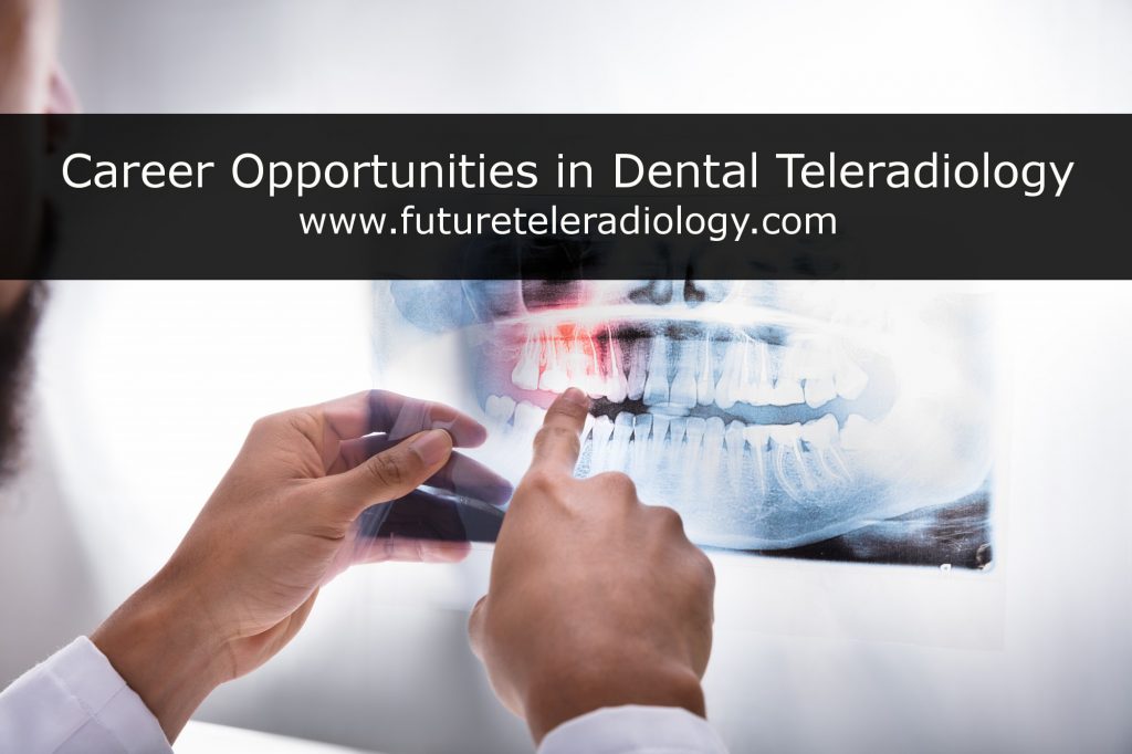
Introduction: Dental radiography plays a pivotal role in modern dentistry, enabling practitioners to diagnose and treat various oral health conditions. However, the field of dental radiography has undergone significant advancements, along with its own set of challenges and triumphs. In this blog, we will explore some of the key aspects of dental radiography, the challenges faced, and the remarkable triumphs in imaging techniques that have transformed the way we approach oral healthcare.
Challenges in Dental Radiography:
- Radiation Exposure: One of the most significant challenges in dental radiography is minimizing radiation exposure to patients. Traditional X-ray machines expose patients to small doses of ionizing radiation, which can accumulate over time. This challenge has spurred the development of digital radiography techniques, such as digital sensors and panoramic radiography, which significantly reduce radiation exposure while maintaining diagnostic accuracy.
- Image Quality: Achieving high-quality images is essential for accurate diagnoses. However, capturing clear images in a small and often confined oral space can be challenging. The development of high-resolution digital sensors and cone-beam computed tomography (CBCT) technology has greatly improved image quality in dental radiography.
- Patient Comfort: Many patients experience anxiety or discomfort during dental radiography procedures. Traditional bite-wing X-rays, for example, involve uncomfortable positioning of film or sensors. Advanced imaging techniques, such as CBCT and intraoral scanners, have made the process more comfortable for patients, reducing anxiety and improving overall patient experience.
Triumphs in Dental Radiography:
- Digital Radiography: The transition from traditional film-based radiography to digital systems has been a game-changer. Digital radiography offers numerous advantages, including instant image acquisition, enhanced image manipulation, and a significant reduction in radiation exposure. This shift has not only improved diagnostic capabilities but also streamlined the workflow in dental practices.
- Cone-Beam Computed Tomography (CBCT): CBCT has revolutionized 3D imaging in dentistry. It provides high-resolution, three-dimensional images of the oral and maxillofacial region. This technology is particularly valuable for implant planning, orthodontics, and diagnosing complex conditions like impacted teeth or temporomandibular joint disorders.
- Intraoral Scanners: Intraoral scanners have made the traditional dental impression process almost obsolete. These handheld devices allow for the quick, precise, and comfortable scanning of a patient’s teeth and oral structures. This not only improves the accuracy of restorations but also enhances the patient experience.
- Artificial Intelligence (AI): AI has been integrated into dental radiography to aid in diagnosis and treatment planning. AI algorithms can quickly analyze images, detect anomalies, and assist dentists in making more informed decisions. This technology holds the potential to further improve the accuracy and efficiency of dental care.
Conclusion:
Dental radiography has come a long way, overcoming significant challenges and achieving remarkable triumphs in imaging techniques. The shift from traditional film-based systems to digital radiography, the introduction of CBCT and intraoral scanners, and the integration of AI have all contributed to a more efficient, accurate, and patient-friendly dental radiography landscape. As technology continues to evolve, the future of dental imaging looks promising, with the potential to further enhance oral healthcare and patient outcomes.
Service Areas:- Surendranagar – Chuda, Chotila, Dhrangadhra, Lakhtar, Limbdi, Muli, Patdi, Sayla, Thangadh, Wadhwan; Ahmedabad – Ahmadabad City, Daskroi, Dholka, Sanand, Viramgam, Bavla, Dhandhuka, Ranpur, Detroj-Rampura, Barwala, Mandal; Vadodara – Savli, Vadodara-Rural, Waghodia, Dabhoi, Padra, Karjan, Shinor, Desar, Vadodara City-East, Vadodara City-West, Vadodara City-North, Vadodara City-South.
